




HistoAtlas
Like
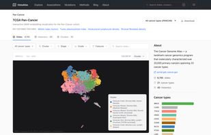
HistoAtlas is a comprehensive resource mapping the morphological landscape of cancer across 21 TCGA cancer types. By extracting quantitative histological features from over 6,000 diagnostic whole-slide images, HistoAtlas reveals how tissue architecture relates to patient...

Cost / License
- Free
- Open Source
 EU
EUPlatforms
- Online


Features
HistoAtlas News & Activities
Highlights All activities
Recent activities
 masterchiefm added HistoAtlas as alternative to GenePad
 Fla added HistoAtlas as alternative to VectorBee
- pierreantoine-bannier added HistoAtlas
 pierreantoine-bannier added HistoAtlas as alternative to Serial Cloner, CLC Genomics Workbench, SnapGene Viewer and Genophore


HistoAtlas information
Developed by
 HistoAtlas
 Licensing
Open Source and Free product.Written in
Alternatives
6 alternatives listedBadge
Get an embeddable badge for HistoAtlasSupported Languages


No comments or reviews, maybe you want to be first?